|
Conférence plénière Biologie |
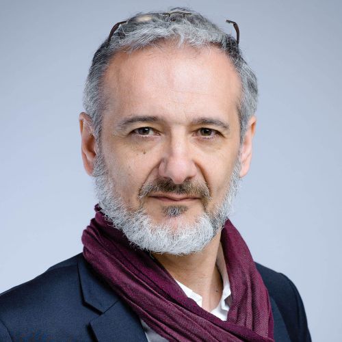
Abstract. Drug discovery is a complex problem, finding a synthetic chemical (Synthetic Modalities) that fulfill all the requirement to become a drug is a challenge. Over the years the process of discovery has evolved from Observation and serendipity to more data-driven and then rationally data driven. Even if we claim from decade that the rational drug design is a core activity of drug discovery the evolution of computing capabilities and the development of new algorithm especially in the field of Artificial Intelligence have given a boost to this domain. Today the Artificial intelligence is under the spotlight in drug discovery and some recent developments in machine learning especially neural networks brought interesting opportunities for the chemical space exploration. In this talk I will present some examples of Machine-learning capabilities, from the single point exploration of chemical space (predictive models) to the optimized exploration of the chemical space provided by the de-novo design by reinforcement learning (GenAI). Machine learning can also be used on top of other more traditional physics-based technologies to increase the capability and access to numbers not usually accessible i.e., Virtual-Screening or combined with Free-Energy Perturbation. Combination of machine learning and first principal technologies gives access to a powerful mixed technology that push the boundaries of research. A lot is expected from of these technologies to explore the “druggability” of some difficult biological systems. Today these algorithms are used daily to support our research portfolio but still the most important operation remain the decision-making process. Although assisted by machine especially using multi-parametric optimization, the human remains at the center. In this respect Artificial Intelligence provides a real improvement of our “In-Silico toolbox” but on a project perspective the intelligence lies into the team. Today we prefer to refer to “Augmented Medchem” or “Augmented Intelligence” when speaking of this shift of ways of working. But still drug discovery remains a complex problem… |
| Conférence plénière Chimie Philippe Boullay CRISMAT, Caen 3D ED : un puissant outil de caractérisation structurale pour la chimie |
Résumé. La cristallographie structurale a connu une révolution au cours des dix dernières années, grâce à l'introduction de divers protocoles d'acquisition et d'analyse de données de diffraction des électrons en faisceaux parallèles. Ces approches, regroupées sous le nom générique de 3D ED, visent toutes à reconstruire le réseau réciproque (3D) d'un composé cristallin en collectant des diagrammes de diffraction des électrons (ED) à différents angles d'inclinaison du goniomètre. Réalisée avec un microscope électronique à transmission, la 3D ED permet d'analyser des monocristaux de taille nanométrique et d'obtenir des données permettant de déterminer la structure cristalline de matériaux à une échelle inaccessible par d'autres techniques de diffraction. Après une introduction à la méthode, dans laquelle nous expliquerons les différentes approches disponibles, nous illustrerons quelques applications de la 3D ED pour différents types d'échantillons et de problématiques. Nous commencerons par la 3D ED assistée par précession, qui permet de résoudre des structures complexes et d'obtenir des affinements fiables et précis pour des composés "stables", typiquement en science des matériaux. Nous parlerons ensuite de la 3D ED en rotation continue, qui permet d'acquérir des données avec des temps de collecte inférieurs à la minute pour des composés "fragiles", typiquement certains matériaux poreux, en pharmacologie et en sciences de la vie. Nous présenterons enfin quelques développements instrumentaux et méthodologiques récents destinés à rendre cet outil encore plus accessible, polyvalent et efficace. |
| Conférence plénière Physique Pauline Martinetto Institut Néel, Grenoble Cristallographie et matériaux du patrimoine culturel : des mesures non invasives sur les œuvres à l’analyse quantitative semi-automatique de phases |
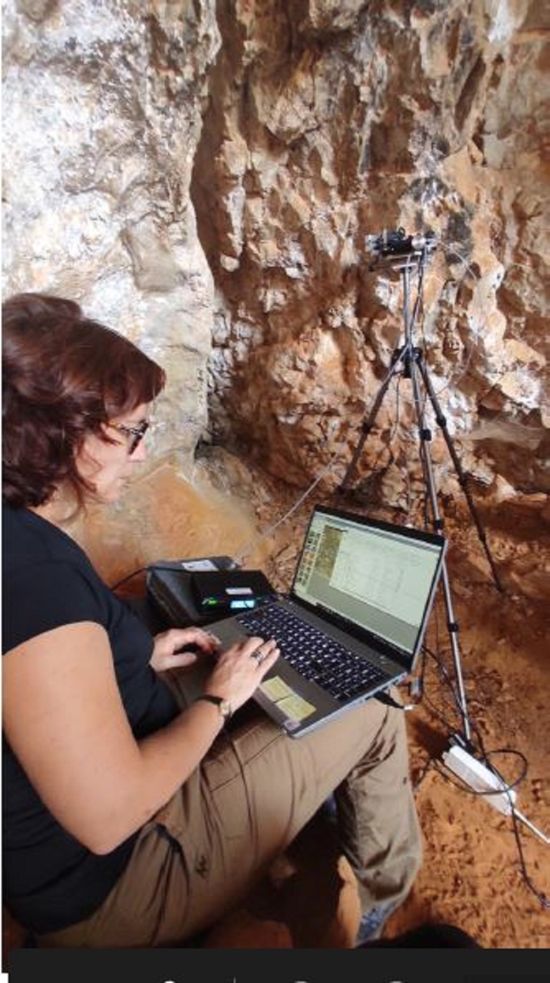
Résumé. Les matériaux du patrimoine culturel sont de plus en plus fréquemment au centre de programmes de recherche interdisciplinaires, dans lesquels les approches des différentes disciplines impliquées sont croisées dans le but de retracer l’histoire des œuvres d’art, de leur création et fabrication jusqu’à leur état de dégradation actuel. A l‘Institut Néel, nous cherchons à développer les méthodes d’analyse utilisant les rayons X afin de révéler les traces enregistrées dans ces matériaux pour identifier les matières premières, leur mélange et si possible leur provenance et les savoir-faire techniques. Dans cet exposé, je présenterai l’approche analytique que nous avons récemment développée, qui combine des mesures non invasives de fluorescence et diffraction des rayons X directement sur les œuvres, et des expériences synchrotron de carto/tomographie de diffraction sur micro-prélèvements. Le recours à ces techniques d’imagerie synchrotron aboutit à l’enregistrement de quantités massives de données qui nous ont poussés à développer des méthodes de traitement semi-automatiques et rapides nous permettant d’obtenir des informations quantitatives en chaque point de l’image, particulièrement informatives pour la compréhension de stratigraphies picturales complexes. J’illustrerai notre approche en donnant différents exemples, issus notamment du programme PATRIMALP (IDEX, UGA), et développerai plus particulièrement l’étude que nous avons menée sur les décors en « brocarts appliqués » d’un corpus de sculptures produites dans les anciens Etats de Savoie à la fin du Moyen Age. |
| L'AFC invite: Andy Maloney - CCDC, Cambridge, UK. With our powers combined… The CSD and its applications across structural science |
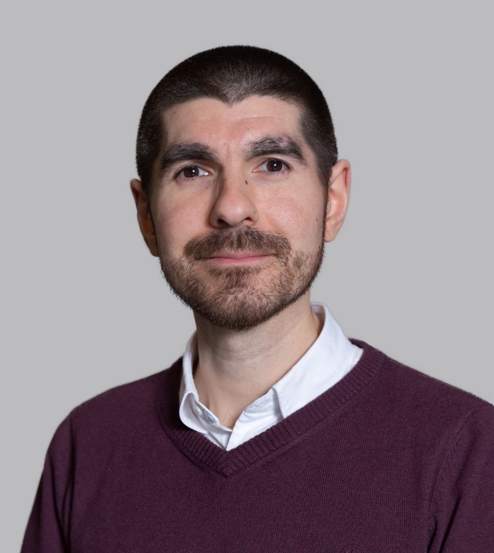
Abstract: Following a vision that the collective use of data would lead to new knowledge and generate new insights Dr Olga Kennard established the Cambridge Structural Database (CSD) in 1965, a step which transformed modern crystallography. Today over one million structures are shared through this resource and the database is something the entire scientific community can be proud of. It is thanks to this community’s exemplary data sharing practices, as well as the thoughts of visionaries in the field, that this collection of data exists. The CSD and the accompanying software are now used by scientists worldwide working in over 70 countries, and the knowledge derived from the structural data contained in the database has underpinned fundamental chemical discoveries and played a key role in designing new materials from drugs to pigments and beyond. This talk will look at some of the research that the CSD has enabled and highlight the knowledge and insights that have been derived from this collection of data. It will explore how the database has evolved over the years and how researchers have used it for a range of different purposes, from rationalising the geometries of macromolecules, to predicting intermolecular interactions, and even to understanding the surface chemistry of crystals themselves. |

 Jean-Philippe Rameau is Senior Distinguished Scientist and head of the “Molecular Design Sciences” department in the Sanofi Research Platform, supporting the Research portfolio in the field of In-Silico Sciences for Rational Data-Driven Drug Discovery. Trained as Computational Chemists he holds a PhD in Physical-Chemistry specialized in Molecular Mechanics and Forces Fields development. He complemented his training with two post- in Pharma on GPCR modeling and in QSPR for Petroleum industry. He joined Sanofi in 1999 as a Research Scientist and took increasing responsibilities in Sanofi-R&D.
 Philippe Boullay a obtenu un doctorat en chimie des matériaux en 1997 à l'Université de Caen où il a travaillé sur la synthèse et la caractérisation structurale d’oxydes de métaux de transition. Il a ensuite été chercheur post-doctorant au département de Chimie Inorganique de l'Université de Stockholm et au laboratoire EMAT de l'Université d'Anvers. En 1999, il devient chercheur CNRS au laboratoire SPCTS de Limoges où il a étudié la structure et les transitions de phase dans les matériaux ferroélectriques. En 2006, il a rejoint le laboratoire CRISMAT à Caen où il a notamment promu et développé l'utilisation de la cristallographie aux électrons pour l'analyse structurale des matériaux fonctionnels.
 Pauline Martinetto a soutenu un doctorat en physique en 2000, effectué en collaboration entre le Centre de Recherche et de Restauration des Musées de France (CNRS, Paris), l’ESRF (Grenoble) et le laboratoire de Cristallographie de Grenoble, sur l’étude cristallographique des préparations cosmétiques de l’Egypte ancienne. Elle est actuellement enseignante-chercheuse à l’Université Grenoble Alpes et effectue sa recherche à l’Institut Néel (CNRS - UGA, Grenoble). Ses projets de recherche consistent à faire évoluer les techniques analytiques utilisant les rayons X, ainsi que les traitements de données associés, afin de les rendre plus pertinents et adaptés à l’étude des matériaux anciens et patrimoniaux. Récemment, dans le cadre de programmes interdisciplinaires à l’interface entre physique, archéologie et histoire de l’art et des techniques, elle a participé au développement d’instruments mobiles pour l’analyse non invasive d’objets et œuvres d’art.
 Andy Maloney has a background in chemical and computational crystallography. He joined the Cambridge Crystallographic Data Centre (CCDC) in 2015, where he now leads the Particle Science Team. Working closely with the pharmaceutical industry, his role involves leading research projects in the field of structure-property relationships. His main interest is in Particle Informatics, developing methods to understand the links between crystal structure, particle properties, and downstream manufacturing processes.